Taipei, Taiwan: Researchers from Academia Sinica and National Taiwan University have developed a novel procedure to mass produce fluorescent nanodiamonds, and have shown the utility of these nanoparticles as biological probes.
Nanodiamonds (NDs) with sizes ranging from around 5nm to over 100nm can be formed by detonation and non-detonation methods, however, NDs require costly post-processing in order to fluoresce. Researchers from Academia Sinica and National Taiwan University have developed a novel procedure to mass produce fluorescent nanodiamonds (FND), with production rates about two orders of magnitude higher than traditional methods. Creating FND traditionally involves the use of a limited-availability van de Graaff accelerator to create a high-energy electron beam followed by annealing at temperatures on the order of 800°C. FNDs created in this way require a high ion dose to create the necessary defects that give the FNDs their fluorescent properties, and thus have not been extensively used despite their suitability for applications such as cellular biomarkers.

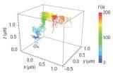
FNDs are suitable for a number of applications, such as biological and medical imaging and spectroscopy, drug delivery, lighting/optical, and other applications requiring nanoscale fluorescent particles. In this research from Taiwan, the FNDs were found to be bright and stable enough that the movement of one FND could be tracked after cellular uptake for over 200 seconds. In other research, FNDs have been shown to be less cytotoxic than other fluorescent nanoparticles and do not photobleach or photoblink like other fluorescent nanoparticles. Non-fluorescent NDs are available commercially in detonation and non-detonation forms and are used for applications such as atomic polishing, polymer additives, lubrication, and composites.
Supplementary material is available from Nature Nanotechnology.
Other pieces about the article:
Images reprinted by permission from Macmillan Publishers Ltd: Nature Nanotechnology, 2008.
